
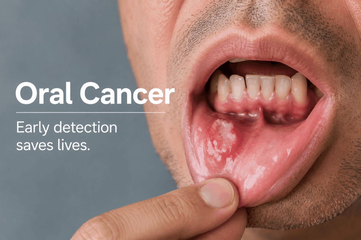
Oral cancer, also called mouth cancer, starts in any part of your oral cavity. That includes your lips, tongue, gums, the floor of your mouth, the roof of your mouth, the inside lining of your cheeks, and the area just behind your wisdom teeth.
It is more common than most people realize. The American Cancer Society estimates that about 60,480 people in the United States will be diagnosed with oral cavity or oropharyngeal cancer in 2026, and roughly 13,150 will die from it.
When oral cancer is found early, before it has spread, treatment is often far simpler than people fear, frequently a single, well-planned procedure rather than a long fight. That gap between early and late detection is the entire reason this guide exists, and it is also the reason a same-day phone call to your dentist is never an overreaction.
Is That a Canker Sore or Oral Cancer? Here Is How to Tell
In the vast majority of cases, what you are dealing with is a canker sore, not cancer.
A canker sore (also called an aphthous ulcer) is a small, shallow, oval-shaped sore with a white or yellowish center and a clearly defined red border. It is painful, often triggered by stress, minor injury, or acidic foods, and it heals completely on its own within 7 to 14 days. It does not bleed unprovoked, and it does not grow.
Oral cancer can start looking similar, but behaves differently over time:
| Feature | Canker Sore | Possible Oral Cancer |
|---|---|---|
| Heals within 2 weeks | Yes | No |
| Painful | Usually yes | Often painless early on |
| Bleeds without being touched | No | Can |
| Borders | Smooth, defined | Irregular, raised |
| Color | White/yellow center, red border | White, red, or mixed patches |
| Grows over time | No | Can |
| Location | Inside lips, cheeks, under tongue | Anywhere in the mouth |
The rule of two weeks is simple and meant to make this easy, not scary: if something in your mouth has not healed within 14 days, get it looked at. Call your dentist, tell them what you are seeing, and let them take it from there.
What Are the Early Signs of Oral Cancer?
The early signs of oral cancer are easy to miss because they often look like ordinary mouth irritation. Most early-stage oral cancer causes no pain at all, which is exactly why so many people ignore it, not because they were careless, but because pain is the signal most of us were taught to watch for.
Here is what to watch for instead:
- A sore or ulcer on your lip or inside your mouth that bleeds easily and has not healed within two weeks
- A white patch (leukoplakia), a red patch (erythroplakia), or a mixed red-and-white patch on your gums, tongue, or inner cheeks that cannot be scraped away
- Rough, crusty, or thickened areas on your lips, gums, or inside your cheeks
- Unexplained bleeding anywhere inside your mouth
- Numbness, pain, or tenderness in your mouth, face, or neck with no obvious cause
- A lump, bump, or thickened area anywhere in or around your mouth or throat
- Swelling in the jaw that makes your dentures sit differently
- Difficulty chewing, swallowing, speaking, or moving your jaw or tongue
- A persistent sore throat or the feeling that something is caught in the back of your throat
- Unexplained weight loss
- Pain in one or both ears
- A hoarse voice that does not resolve
The absence of pain is not reassurance on its own, but having one or two of these on the list is not a diagnosis either. It is simply your cue to get a professional opinion. If you see any of these early signs of oral cancer and they have been present for two weeks or more, call your dentist. That is the whole next step.
What Does Oral Cancer Look Like? (Visual Signs)
People searching for oral cancer pictures want a clear, plain-language description of what they are looking at, so here is exactly what each type looks like.
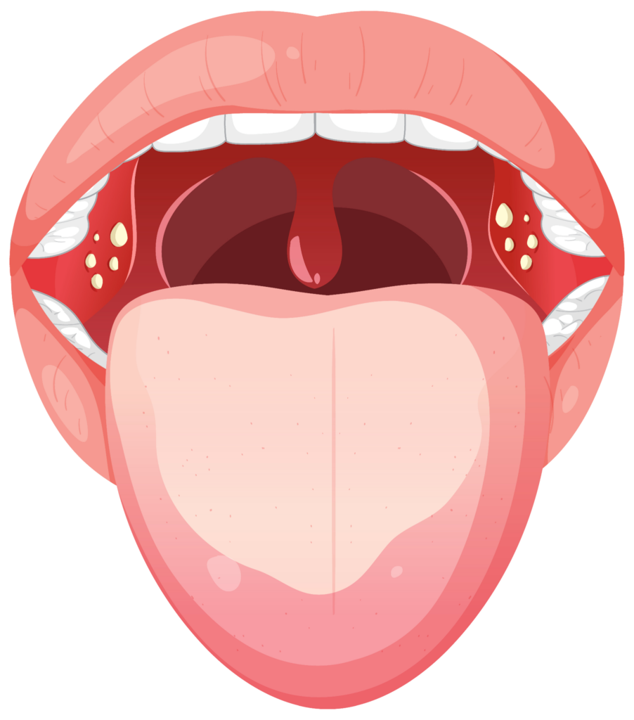 |
White patches (leukoplakia: Flat, white or grayish areas on the inside of the cheek, tongue, or gums. They look almost like a dried film over the tissue. You cannot wipe them away with your finger. They are considered pre-cancerous and should always be evaluated by a dentist or oral surgeon. |
|---|---|
| Red patches (erythroplakia): Bright red, velvety-looking areas anywhere in the mouth. These carry a higher risk of being cancerous than white patches and are taken seriously even at small sizes. | 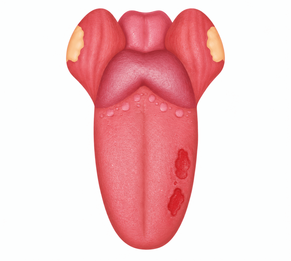 |
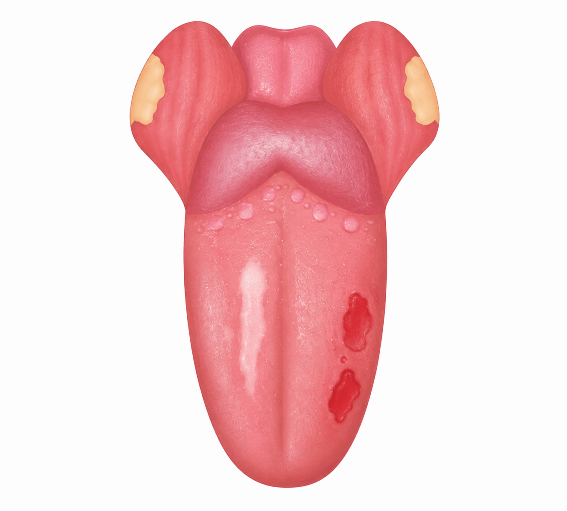 |
Mixed red-and-white patches (erythroleukoplakia): Areas with both colors present together. These mixed patches have a higher malignancy rate than purely white ones. |
| Non-healing sores: Unlike a regular canker sore with smooth, rounded edges that resolves in under two weeks, oral cancer sores persist, bleed easily when touched, and often have raised, uneven, or cratered edges. | 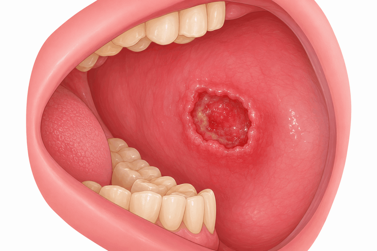 |
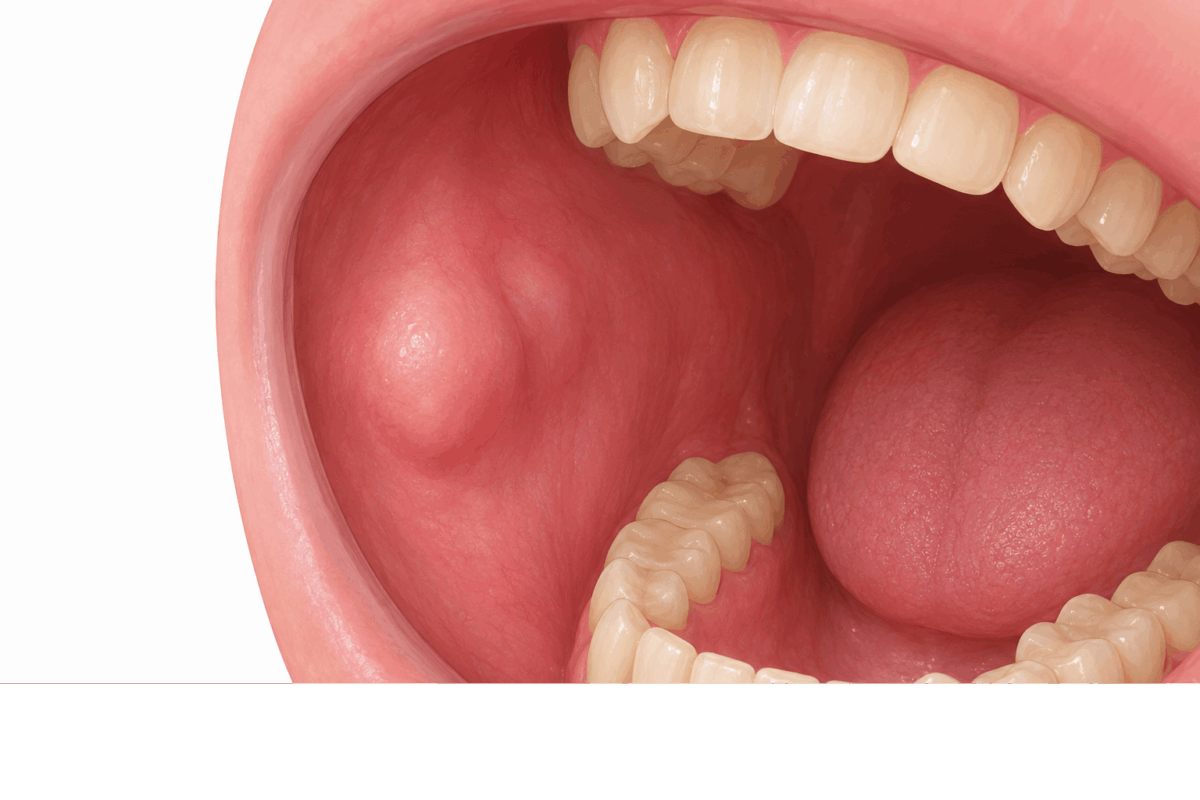 |
Lumps or thickened areas: A hard lump or area of thickening anywhere inside the mouth, on the lip, or along the jaw. These are often completely painless, which makes them easy to dismiss. Worth a look either way. |
| Ulcers with irregular borders: A standard mouth ulcer has a clean, round shape. A suspicious ulcer may look asymmetrical, have uneven raised edges, and resist healing over multiple weeks. | 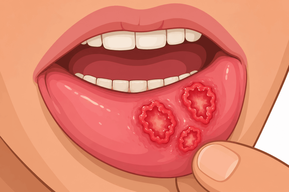 |
Any of these changes that have been present for two weeks or more deserve a professional evaluation. A photograph on your phone is a great way to track changes, but it is not a substitute for a dentist actually examining it.
Where Does Oral Cancer Usually Start?
Oral cancer most commonly starts on the tongue and the floor of the mouth, which is the soft tissue directly underneath your tongue. Together, these two sites account for the majority of all oral cavity cancer cases in the United States.
Other common sites include:
- The lips, particularly the lower lip, due to sun exposure
- The gums
- The inside lining of the cheeks
- The hard palate (the bony front portion of the roof of your mouth)
- The area immediately behind the wisdom teeth (retromolar trigone)
Cancer that starts in the back of the throat, the base of the tongue, the tonsils, or the soft palate is called oropharyngeal cancer and is increasingly linked to HPV. It is treated as a related but separate condition from oral cavity cancer.
What Causes Oral Cancer?
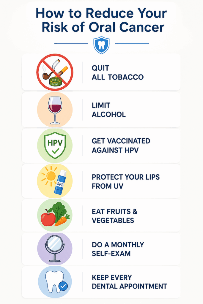
Oral cancer begins when DNA mutations cause normal cells in the mouth to grow and multiply out of control. Several risk factors significantly increase the likelihood of this happening, and knowing them is genuinely useful, both for prevention and for knowing you are not alone in this.
| Risk Factor | Key Point |
|---|---|
| Tobacco Use | Leading cause; smoking, chewing tobacco, and vaping all increase risk. |
| Heavy Alcohol Use | Heavy drinking raises risk, especially when combined with tobacco. |
| HPV | HPV-16 is a major cause, particularly in younger adults. |
| Sun Exposure | UV exposure increases the risk of lip cancer. |
| Age | Most cases occur after age 60, but younger people can also be affected. |
| Poor Nutrition | Low fruit and vegetable intake is linked to higher risk. |
| Family History | Having a family history of oral cancer increases risk. |
Oral Cancer Risk Factors at a Glance
| Risk Factor | Risk Level |
|---|---|
| Tobacco use (any form, including vaping) | Very High |
| Heavy alcohol use | High |
| Tobacco and alcohol combined | Extremely High |
| HPV infection (HPV-16) | High, especially oropharyngeal |
| Age over 55 | Moderate to High |
| Excessive sun exposure (lip cancer) | Moderate |
| Low fruit and vegetable diet | Moderate |
| Family history of oral cancer | Moderate |
| No known risk factors | Still possible |
Oral Cancer Stages: What They Mean for Your Treatment
Oral cancer is staged from 0 to IV based on tumor size and how far the cancer has spread. The stage drives every treatment decision, and the short version of this whole section is the most encouraging fact in this entire article: the earlier it is caught, the simpler treatment usually is.
- Stage 0: Abnormal cells are confined to the surface lining of the mouth. Highly treatable, often resolved with minor surgical removal.
- Stage I: A small tumor (2 cm or smaller) that has not spread to lymph nodes. Excellent outcomes, and frequently treated with surgery alone.
- Stage II: Tumor has grown between 2 and 4 cm but has not reached the lymph nodes. Still very treatable with surgery and sometimes radiation.
- Stage III: Tumor is larger than 4 cm, or cancer has spread to one lymph node on the same side as the tumor. Treatment typically combines surgery, radiation, and sometimes chemotherapy.
- Stage IV: Cancer has spread to nearby structures, multiple lymph nodes, or distant organs. Treatment is more involved at this stage, which is precisely why catching things earlier changes the entire path forward.
What Happens If Oral Cancer Goes Untreated?
Oral cancer does not stay in one place. Left untreated, it spreads from the original site to surrounding tissue, then to lymph nodes in the neck, and eventually to distant organs. As it progresses, symptoms become more difficult to manage, including pain, trouble swallowing, and weight loss.
Every stage of progression narrows your options. Catching something early is genuinely the easiest version of this entire process, and most people who get checked promptly never end up anywhere near this part of the article. That is the outcome we are working toward with you.
How to Check for Oral Cancer at Home (Step-by-Step Self-Exam)
A monthly self-exam takes less than five minutes and only needs a mirror and good light. Here is exactly how to do it.
Step 1: Lips
Pull your upper and lower lips away from your teeth and look at the inside surface. Feel along both lips with your fingers. Look for any sores, patches, lumps, or color changes.
Step 2: Gums
Check your upper and lower gums all the way around. Look for red or white patches, swelling, or sores.
Step 3: Cheeks
Use your fingers to pull each cheek out gently. Look at the inner lining. Feel it for any lumps or thickened areas. Check for any red, white, or mixed-color patches.
Step 4: Tongue
Stick your tongue out and look at the top surface. Then use your fingers to gently hold your tongue and look at the sides and the underside. The sides of the tongue are one of the most common sites for oral cancer. Feel along the tongue for any lumps or hardened areas.
Step 5: Floor of the mouth
Lift your tongue to the roof of your mouth and look at the tissue underneath. This is the other most common site for oral cancer. Feel it with your fingertip.
Step 6: Roof of the mouth
Tilt your head back and open wide. Look at the hard palate (the bony front part) and the soft palate (the softer back portion). Look for any color changes, sores, or lumps.
Step 7: Throat and neck Using both hands, feel along the sides of your neck, your jaw line, and under your chin for any swollen lumps or enlarged lymph nodes that feel hard or do not move when pressed.
Most months, you will find nothing, and that is exactly the point of doing this regularly: it teaches you what normal looks and feels like for you, so anything new actually stands out. If you do find something that was not there last month, or anything that has not changed or healed in two weeks, call your dentist and document it with a phone photo so you can compare it at your appointment
How Is Oral Cancer Diagnosed?
A proper oral cancer diagnosis requires a clinical examination and, if anything suspicious is found, a biopsy. Here is how the process works, step by step, so there are no surprises.
Visual and physical exam
Your dentist examines your entire mouth, lips, tongue, gums, cheeks, throat, jaw, and neck. They look and feel for any abnormal tissue, patches, sores, or lumps.
Brush biopsy (exfoliative cytology)
A small brush or spatula collects surface cells from a suspicious area painlessly. The cells are sent to a lab for analysis.
Incisional biopsy
If a lesion looks suspicious or a brush biopsy returns abnormal results, a small piece of tissue is removed under local anesthetic for full pathological analysis. This is the only way to confirm or rule out oral cancer definitively, and it is a quick, well-tolerated procedure.
Imaging
Once cancer is confirmed, CT scans, MRIs, or PET scans map how far it has spread, which determines staging and guides treatment planning.
Specialist referral
Depending on findings, you may be referred to an oral and maxillofacial surgeon, an ENT specialist, or a head and neck oncologist for further evaluation and treatment. We coordinate this for you so you are never left figuring out next steps on your own.
How Is Oral Cancer Treated?
Treatment depends on the stage, location, and your overall health, and most plans involve one or more of the following approaches. The good news woven through all of this: the earlier the stage, the more likely treatment is straightforward.
Surgery
Surgery is the most common first-line treatment for oral cavity cancer. The surgeon removes the tumor along with a margin of healthy surrounding tissue to ensure clean edges. For larger tumors, nearby lymph nodes may also be removed in a procedure called a neck dissection.
When surgery removes significant tissue from the tongue, jaw, or floor of the mouth, reconstructive surgery using tissue grafts or bone reconstruction restores function and appearance.
Radiation Therapy
Radiation uses high-energy beams to destroy oral cancer cells. It can be used alone for small, early-stage tumors, or combined with surgery and chemotherapy for more advanced cases. Dental care before and during radiation matters because radiation to the mouth and jaw can cause dry mouth and tooth decay that are far easier to manage proactively than after the fact.
Chemotherapy
Chemotherapy drugs kill cancer cells throughout the body and are typically paired with radiation rather than used alone for mouth cancer. The combination makes cancer cells more sensitive to radiation and improves overall treatment outcomes.
Targeted Therapy
Targeted therapy drugs work against specific proteins on oral cancer cells that drive their growth. Cetuximab (Erbitux) is one FDA-approved targeted therapy used for head and neck cancers, including oral cancer, particularly when standard chemotherapy is not appropriate.
Immunotherapy
Immunotherapy activates your immune system to recognize and destroy oral cancer cells. It is increasingly used for advanced or recurrent cases, particularly when other treatments have not been effective. Pembrolizumab (Keytruda) is one immunotherapy drug FDA-approved for recurrent or metastatic head and neck squamous cell carcinoma.
Palliative Care
For advanced oral cancer where cure is not the goal, palliative care focuses on symptom management, pain relief, nutritional support, and quality of life. It can be provided alongside active cancer treatment at any stage.
Oral Cancer Screening in Colorado Springs, CO
At Robison Associates in Colorado Springs, our oral cancer screening is a quick, painless, non-invasive exam built into your regular dental checkup. Dr. Letha Robison and our team examine your mouth, tongue, gums, throat, and lymph nodes thoroughly, and use advanced fluorescence technology to catch tissue changes a visual exam alone can miss.
If we find anything that concerns us, we walk you through the next steps clearly and calmly. You will never leave our office wondering what happens next.
We recommend oral cancer screening every six months. If you use tobacco, drink heavily, have a history of HPV, or are over 55, consistent screening matters even more, but it is genuinely valuable for every adult.
Schedule your screening today. Catching oral cancer early is not luck. It is a decision you make at your next dental appointment, and we will be glad to see you.